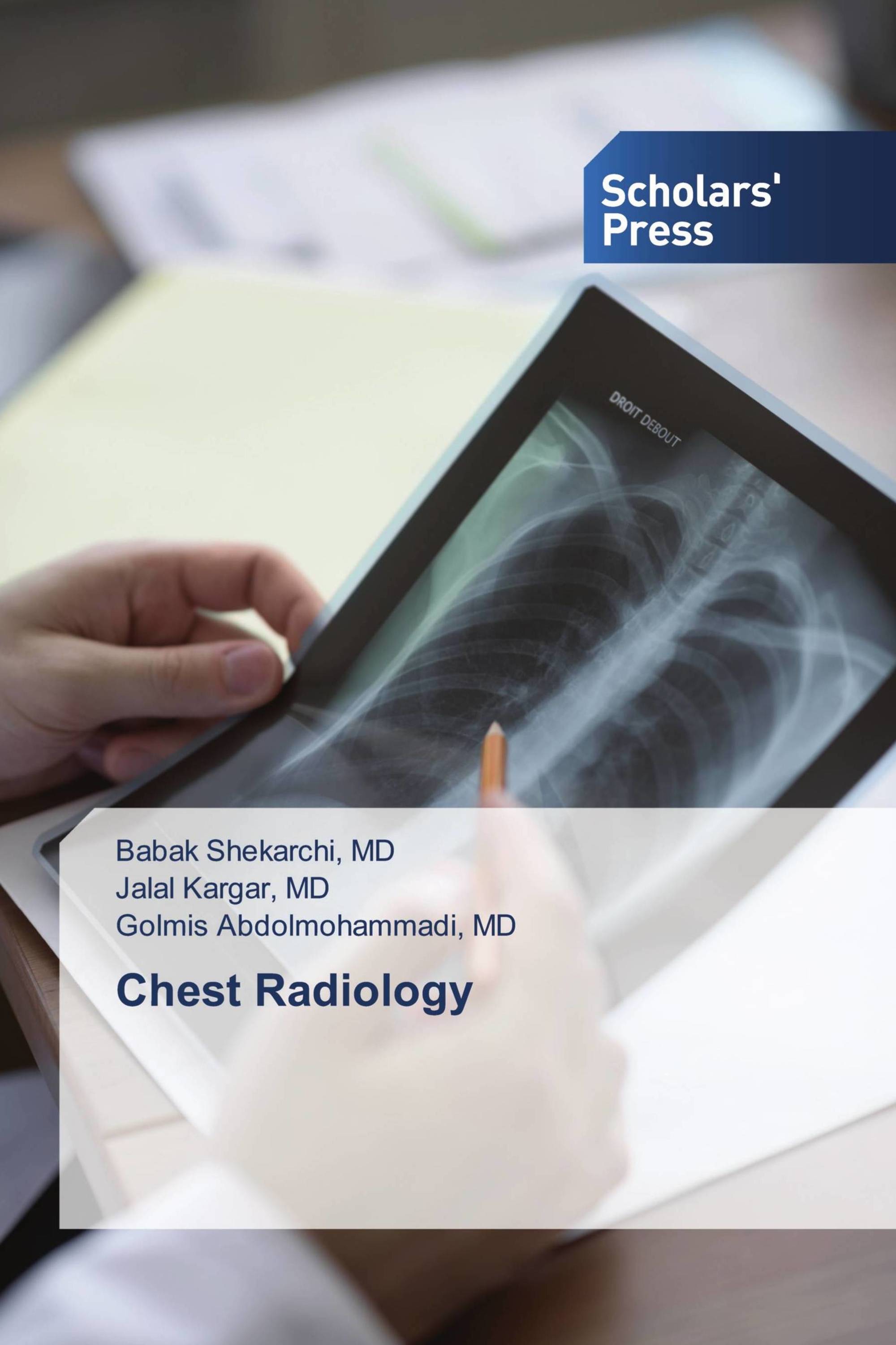
The thorax is the upper part of the human body and is a collection of bone, muscle, blood vessels, and the nervous system. The heart and lungs are two vital organs located in this part of the body and behind the ribs. The head and upper neck, the upper limbs on both sides, and the digestive organs are located below the chest. The bony part of the chest anatomy consists of the sternum, ribs, and thoracic vertebrae. The sternum is one of the broad bones of the human skeleton located in the midline of the chest. This T-shaped bone is one of the protective structures of the internal organs of the chest. This bone is composed of three parts: the manubrium, the body, and the xiphoid process, which are connected by cartilage in childhood, but in adulthood, the cartilage turns into bone and the single-piece sternum forms part of the chest anatomy. This book has chapters including: Chapter I, Chest X-ray basic interpretation; Chapter II, HRCT basic interpretation; Chapter III, CT coronary angiography basic interpretation; Chapter IV, Cardiac MR Basic Interpretation; Chapter V, Ultrasound of lung and diaphragm; References.
Détails du livre: |
|
|
ISBN-13: |
978-3-639-66371-6 |
|
ISBN-10: |
3639663713 |
|
EAN: |
9783639663716 |
|
Langue du Livre: |
English |
|
de (auteur) : |
MD, Babak Shekarchi |
|
Nombre de pages: |
104 |
|
Publié le: |
15.01.2025 |
|
Catégorie: |
Médecine |


